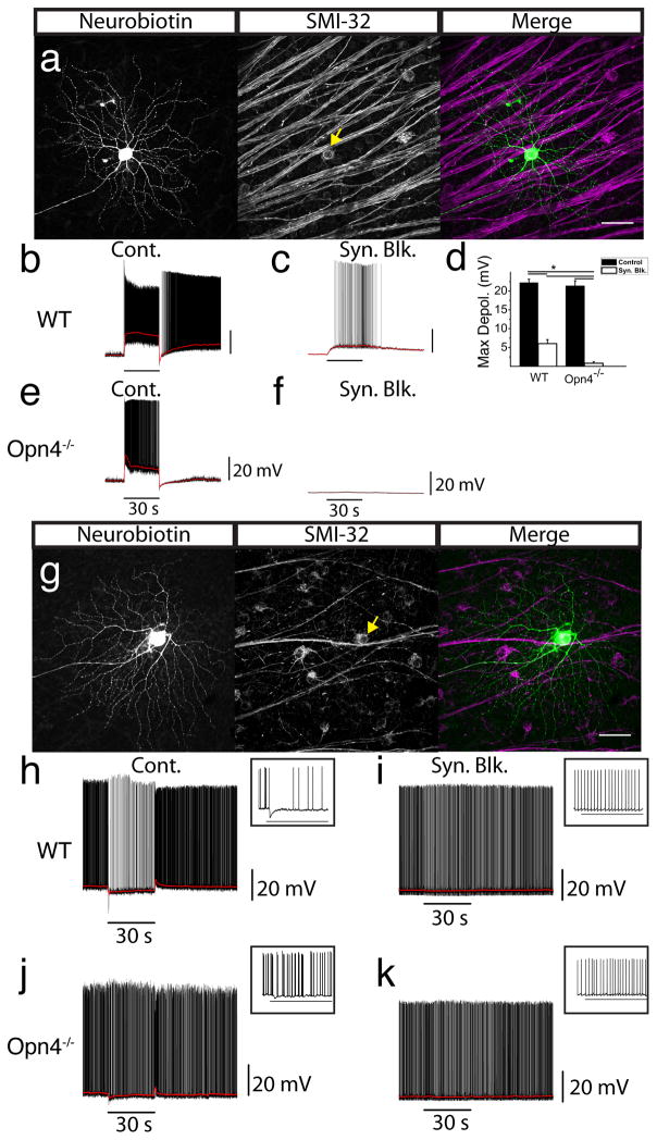
Fig. 2. ON, but not OFF, alpha cells are intrinsically photosensitive.
Cells with the largest soma sizes were randomly targeted for whole cell recordings. (a) Cell recorded in panel b filled with Neurobiotin (left panel) and coimmunostained for SMI-32 (middle panel). Merged image (right panel) shows colocalization of Neurobiotin (green) and SMI-32 (magenta). (b) Whole cell current clamp recording of light response from ON alpha RGC in WT retina. (c) Cell from b recorded in the presence of a cocktail of synaptic blockers. (d) Mean ± SEM maximum depolarization in ON alpha (SMI-32+) RGCs from WT (n = 8) and Opn4−/− (n = 5) mice under control conditions (black bars) and then in the presence of synaptic blockers (white bars). (e) Whole cell current clamp recording from ON alpha RGC in Opn4−/− retina. (f) Cell from e recorded in the presence of synaptic blockers. (g) Cell recorded from in panel h filled with Neurobiotin (left panel) and coimmunostained for SMI-32 (middle panel). Merged image (right panel) shows colocalization of Neurobiotin (green) and SMI-32 (magenta). (h) Whole cell current clamp recording from OFF alpha RGC in a WT mouse. (i) Cell from h recorded in the presence of a cocktail of synaptic blockers. (j) Whole cell current clamp recording from OFF alpha RGC in Opn4−/− retina. (k) Cell from j recorded in the presence of synaptic blockers. (a,g) Scale bars 50 μm. (b–e, h–k) All cells were stimulated with 30s, full-field, 480 nm stimulus. Horizontal bars indicate 30 s light stimulus. (h–k) Inset shows first ~3.5s of response following light onset. * indicates p < 0.01. RGC: retinal ganglion cell.

