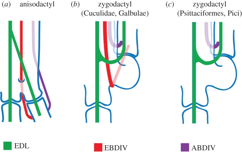
Figure 6.

The topology of dIV tendons. (a) Anisodactyl, (b) zygodactyl with EBDIV and (c) zygodactyl lacking the EBDIV. In (b), the EBDIV does not pass through the canalis interosseus distalis. In (c), the tendon is absent. Modified from [11]. (Online version in colour.)
