Figure 5.
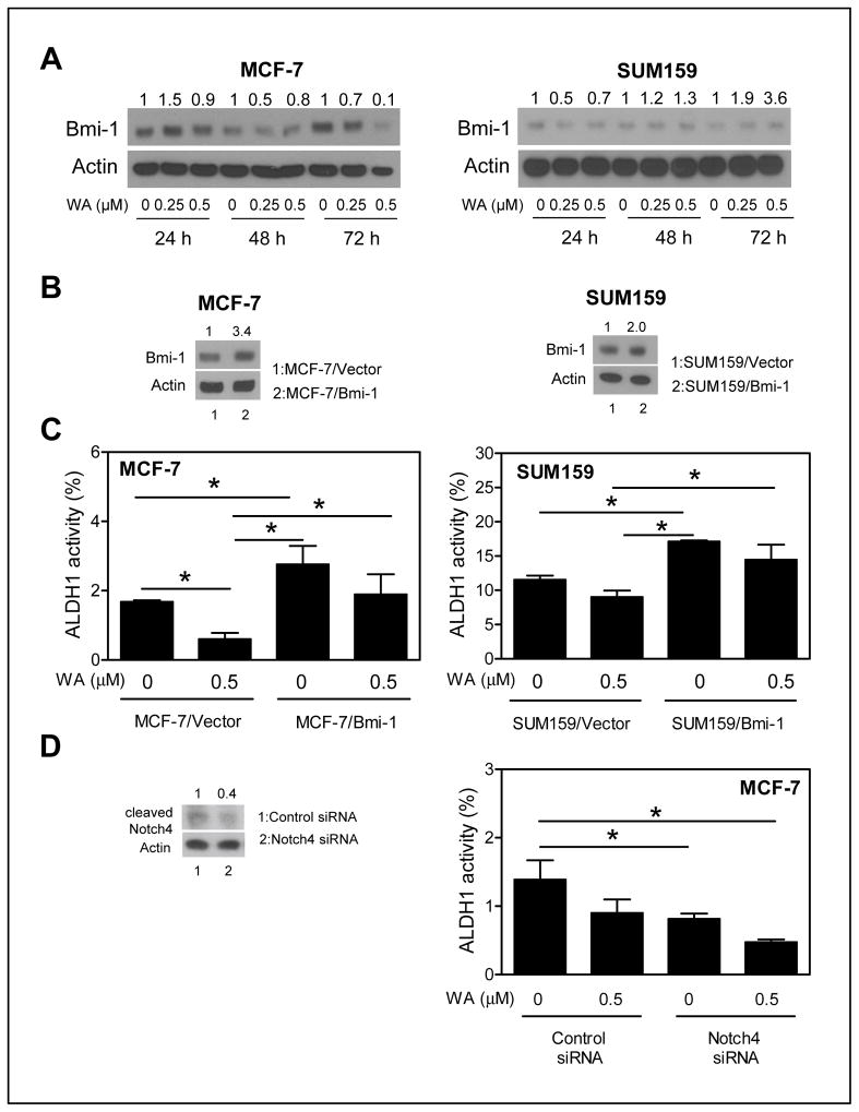
Ectopic expression of Bmi-1 conferred partial protection against ALDH1 activity inhibition by WA. A, western blotting for Bmi-1 protein using cell lysates from MCF-7 and SUM159 cells after treatment with DMSO or WA. Densitometric quantitation relative to respective DMSO control and after normalization for protein loading (actin) is shown above the band. B, western blotting for protein levels of Bmi-1 in MCF-7 (stable transfection) or SUM159 cells (transient transfection) transfected with empty vector (lane 1) or Bmi-1 plasmid (lane 2). C, percentage of ALDH1 activity in empty vector transfected or Bmi-1 overexpressing MCF-7 and SUM159 cells after 72 h (MCF-7) or 24 h (SUM159) treatment with DMSO or 0.5 μmol/L WA. The results shown are mean ± SD (n = 3). *Significantly different (P < 0.05) between the indicated groups by one-way ANOVA with Tukey’s post-hoc analysis. D, ALDH1 activity in MCF-7 cells transfected with a control siRNA or a Notch4 targeted siRNA after 48 h treatment with DMSO or 0.5 μmol/L WA. The western blot shows knockdown of cleaved Notch4. The results shown are mean ± SD (n = 3). *Significantly different (P < 0.05) between the indicated groups by one-way ANOVA followed by Bonferroni’s multiple comparison test. Comparable results were observed in two independent experiments

