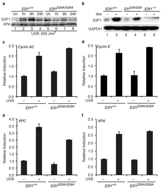
Figure 2. E2F1 stabilization and target expression in response to DNA damage in E2f1S29A/S29A cells.
(a) Primary keratinocytes were isolated from wild type (E2f1+/+, lanes 1-4) and E2f1S29A/S29A (lanes 5-8) mice and left untreated (lanes 1 and 5) or exposed to 250 J/m2 of UVB and harvested at the time points indicated (lanes 2-4 and 6-8). Western blot analysis was performed for E2F1 and XPA. b) MEFs were isolated from wild type (E2f1+/+, lanes 1 and 2), E2f1S29A/S29A (lanes 3 and 4) or E2f1-/- mice and left untreated (lanes 1, 3, and 5) or exposed to 1μM of doxorubicin for 24 h (lanes 2, 4, and 6). Western blot analysis was performed for E2F1 and GAPDH. (c-f) Primary keratinocytes isolated from wild type (E2f1+/+) and E2f1S29A/S29A mice were untreated or exposed to 250 J/m2 of UVB and harvested 8 h post irradiation. Real-time PCR was used to examine expression levels of (c) cyclin A2, (d) cyclin E and (e) XPC, and (f) XPA. The average of two independent samples per group is presented.

