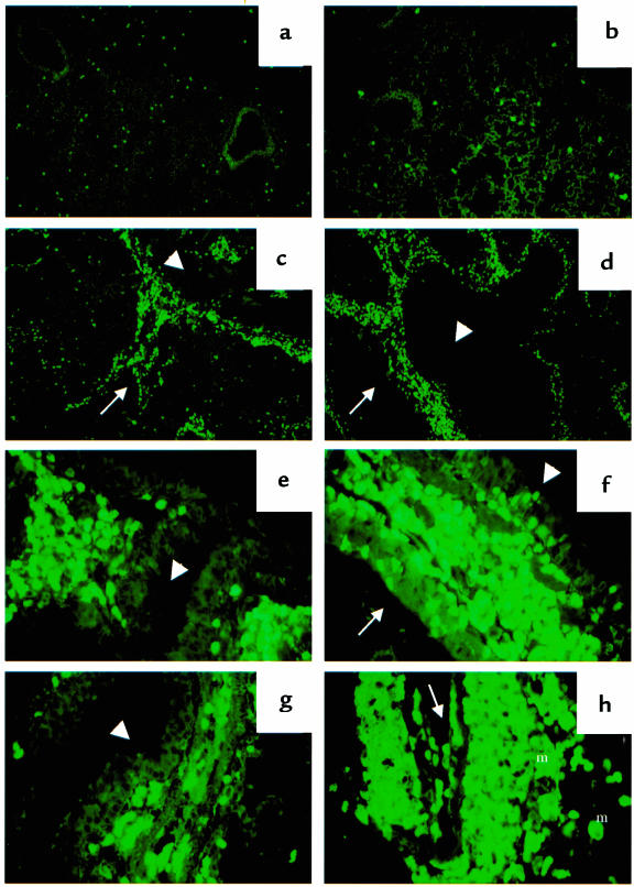
Figure 5.
Immunostaining of lung tissue for mouse eosinophil MBP. H-2Aβ0, HLA-DQ6, or HLA-DQ8 mice were immunized, boosted, and challenged intranasally with SRW allergenic extract as described in Methods. PBS-treated, PBS-challenged mice served as a control for this study. (a) Lung of PSB-treated HLA-DQ6 mouse with a few eosinophils in lung tissue (×10). (b) Lung of SRW-sensitized and -challenged H-2Aβ0 mouse (×10). Severe perivascular and peribronchial concentration of eosinophils in SRW-treated HLA-DQ6 (c) and HLA-DQ8 (d) mice (×10). (e–h) High-power magnification of lungs from HLA-DQ mice (×40). Eosinophil migration from blood vessel to airway mucosa and epithelium in HLA-DQ6 (e) and HLA-DQ8 (f) transgenic mice, and their granule release (f and g). Positive staining of infiltrating macrophages for MBP (h) as a result of its uptake from degranulating eosinophils. Arrow, blood vessel; arrowhead, bronchiole; m, macrophages.

