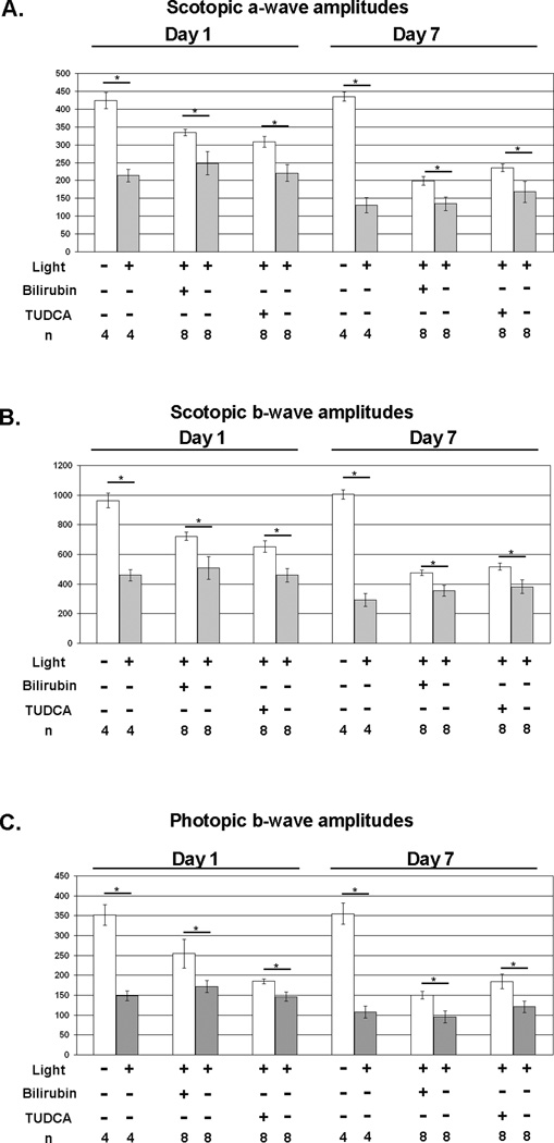
Figure 1. Bilirubin and TUDCA reduce loss of retinal function due to constant light exposure in BALB/c mice.
BALB/c mice were given subcutaneous injections of 5 mg/kg of bilirubin, 500 mg/kg of TUDCA, or corresponding vehicle 24 and 1 hour before constant exposure to 5,000 lux of white light for 8 hours and then returned to a 12 hour/12 hour normal intensity light/dark cycle. Some mice were not exposed to constant light to serve as controls. Scotopic and photopic electroretinograms (ERGs) were done 1 and 7 days after light exposure. The bars show mean (±SEM) scotopic a-wave (A) and b-wave (B) amplitudes recorded after a flash intensity of 25 cd·sec/m2 and mean (±SEM) photopic b-wave amplitudes (C) recorded after a flash intensity of 25 cd·sec/m2 with a background light intensity of 30 cd·sec/m2. Compared to their corresponding vehicle (HSA or NaHCO3) controls, mice treated with bilirubin or TUDCA had significantly higher scotopic a-wave (A), scotopic b-wave (B), and photopic b-wave amplitudes at 1 and 7 days after light exposure.
*p<0.05 by unpaired student t-test for difference between treated mice exposed to light toxicity and the respective vehicle-treated control exposed to light toxicity.

