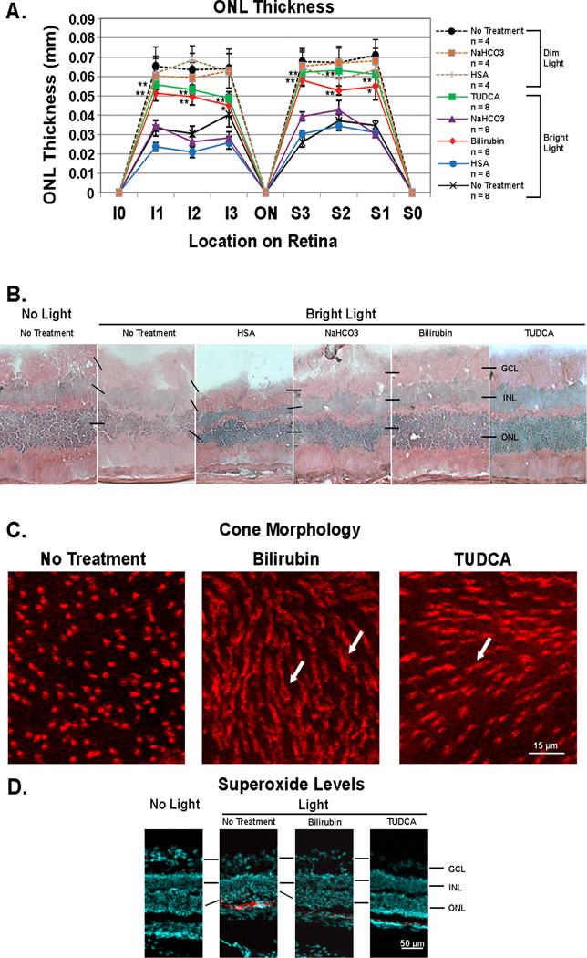
Figure 2. Bilirubin and TUDCA reduce rod cell death, loss of cone outer segments, and accumulation of superoxide radicals after constant light exposure in BALB/c mice.
Twenty-four and again one hour prior to 8 hours of constant exposure to 5,000 lux of white light, BALB/c mice were given subcutaneous injections of bilirubin (5 mg/kg) or TUDCA (500 mg/kg), or no injections and then returned to a 12 hour/12 hour normal intensity light/dark cycle. For comparison, dim-light control mice were given each of the above treatments or no treatment and maintained in normal cyclic light. After 7 days some mice were euthanized and eyes were used for measurement of outer nuclear layer thickness (ONL) on serial ocular sections or retinas were dissected intact and stained with rhodamine-labeled peanut agglutinin (PNA) to visualize cones. Other mice were given two intraperitoneal injections of hydroethidine (20 mg/kg) 30 minutes apart 18 hours prior to death so that superoxide radicals could be visualized in ocular sections. (A) The points show mean (±SEM) ONL thickness at three locations (I1, I2, and I3) between the inferior border of the retina at 6:00 (I0) and the optic nerve (ON) and at three locations (S1, S2, and S3) between the superior border of the retina at 12:00 (S0) and the ON. At all six locations, the mean ONL thickness was significantly greater for bilirubin-treated or TUDCA-treated mice compared to respective vehicle-treated (HSA or NaHCO3) mice (*p<0.05; **p<0.01 by unpaired student t-test for difference from respective vehicle-treated control). There was no difference among dim-light controls including mice treated with bilirubin or TUDCA for which data are not shown. (B) Representative sections from the S1 location show thinning of the ONL and vacuoles in the inner retina of light-exposed mice given no treatment or treated with human serum albumin (HSA) or NaHCO3. In contrast light-exposed mice treated with bilirubin or TUDCA showed thick ONLs and less disruption of the retina. (C) Fluorescence microscopy of retinal flat mounts 0.5 mm superior to the optic nerve showed loss of outer segments and flattening of inner segments of cones in untreated light-exposed mice, whereas inner and outer segments were preserved in bilirubin- and TUDCA-treated mice. Observations were made in three mice from each group and these images are representative. (D) Eighteen hours after injection of hydroethidine, there was strong fluorescence indicating the presence of superoxide radicals in the outer retina of untreated light-exposed mice, but none in the retinas of bilirubin- or TUDCA-treated light-exposed mice or mice that were not exposed to constant light (no light).

