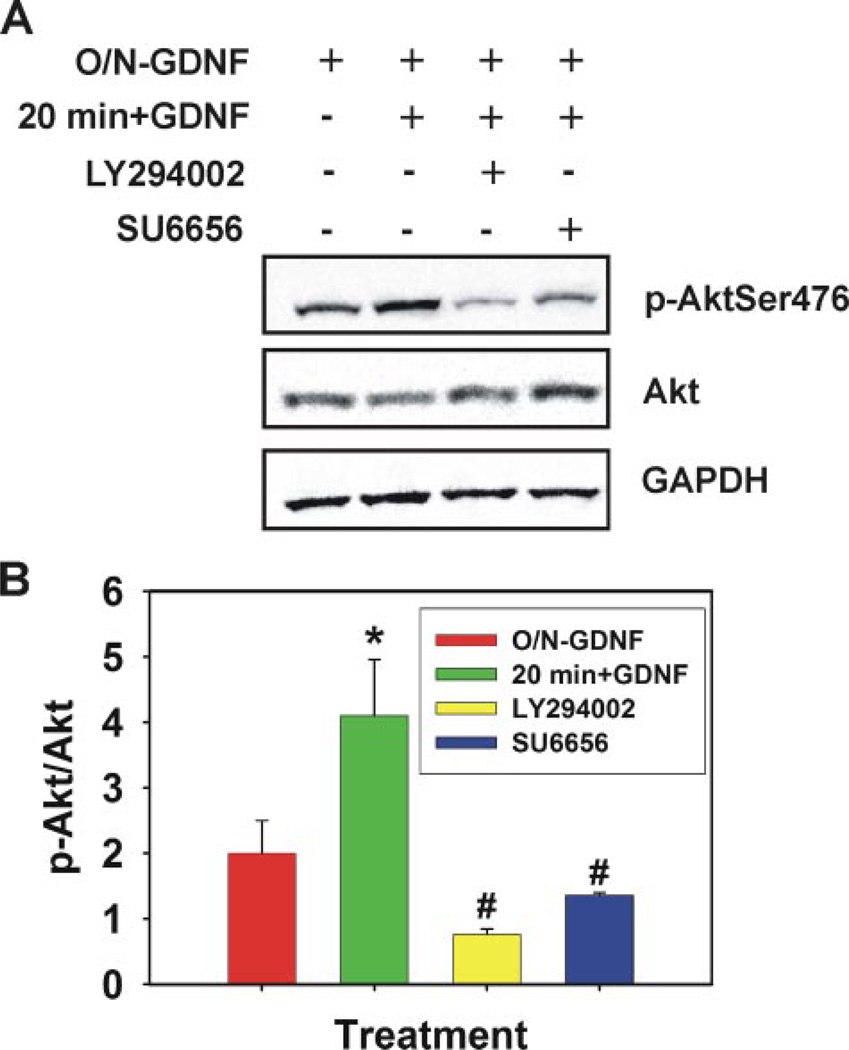
FIGURE 3. GDNF activation of Akt in cultured self-renewing SSCs.
A, representative image of immunoblot analysis for phosphorylated Ser476 Akt levels in cultured self-renewing SSCs. GDNF was removed from culture media overnight for 18 h (O/N-GDNF) followed by a 30-min preincubation with Me2SO (control), 10 µm LY294002 (PI3K specific inhibitor), or 1 µm SU6656 (selective SFK inhibitor). GDNF was then replaced in the media for 20 min before cell lysis. Fifty µg of total cell lysate was resolved by SDS-PAGE, transferred to nitrocellulose membranes, and probed with a phosphorylated Ser-476 Akt-specific antibody. Blots were stripped and re-probed for total Akt and glyceraldehyde-3-phosphate dehydrogenase (GAPDH). B, graphic representation of the average density ratio of phosphorylated (p) Ser-476 Akt to total Akt from three independent immunoblot experiments. Data are presented as the mean ± S.E. The asterisk denotes significant difference (p = 0.024) between O/N-GDNF and 20 min + GDNF. The number symbol (#) denotes significant difference between 20 min + GDNF and LY294002 (p = 0.002) and SU6656 (p = 0.003).

