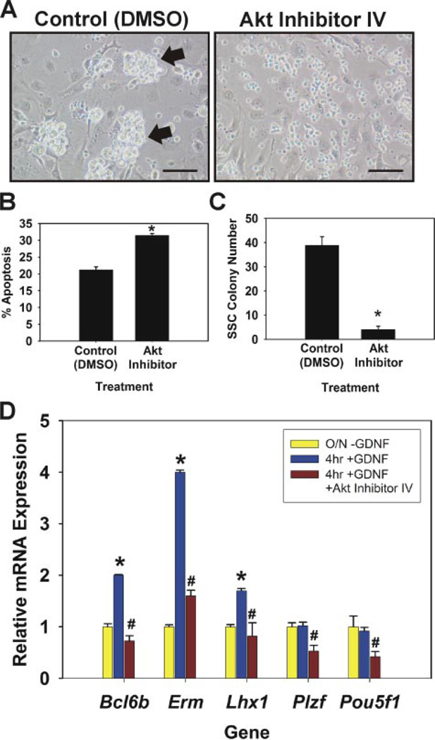
FIGURE 4. Evaluation of Akt inhibition on self-renewing SSCs in vitro.
A, morphological observation of germ cell clump formation 7 days after inclusion of Me2SO (DMSO) or 20 nm Akt inhibitor IV in culture media. In control Me2SO cultures large robust germ cell clumps formed (arrows), whereas only single cells were present in Akt inhibitor cultures. Scale bars, 100 µm. B, percentage of apoptotic cells in clump-forming germ cell cultures 24 h after addition of Me2SO (control) or 20 nm Akt inhibitor IV to culture media. C, functional transplantation analysis of Akt inhibitor treatment on SSC maintenance in vitro. Clump-forming germ cells were cultured for 7 days in conditions that support SSC self-renewal with the addition of Me2SO or 20 nm Akt inhibitor IV. Cells were subsequently transplanted into recipient testes to determine the number of SSCs present in each culture. SSC colony number is the number of colonies within recipient testes/105 cells cultured. Data are presented as the mean ± S.E. for two independent experiments. Asterisks denote significant difference from control. D, qRT-PCR analysis of Akt inhibition on GDNF-regulated gene expression. Cultured SSCs were deprived of GDNF for 18 h (O/N-GDNF, yellow bars) followed by 4 h GDNF replacement (4 h + GDNF, blue bars) with the addition of Me2SO (control) or 20 nm Akt inhibitor IV (red bars). Relative mRNA expression levels for each gene were normalized to that of ribosomal protein S2 and are presented as -fold change compared with O/N-GDNF (yellow bars). Data are the mean ± S.E. for three independent experiments. The asterisk denotes significant difference (p = 0.05) between O/N-GDNF and 4 h + GDNF. The number symbol (#) denotes significant difference (p ≤ 0.01) between 4 h + GDNF and 4 h + GDNF + Akt Inhibitor IV.

