Figure 2.
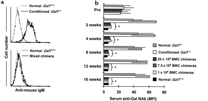
Reduced anti-Gal NAb levels in sera of GalT+/+→GalT–/– mixed chimeras. (a) Representative histograms obtained by FCM analysis show an absence of anti-Gal NAb’s in GalT+/+→GalT–/– mixed chimeras. C.B.-17 scid mouse (GalT+/+) spleen and BMCs were stained with sera from normal GalT+/+, from control conditioned GalT–/– mice, or from BMT recipients; NAb’s were detected using rat anti-mouse IgM-FITC as secondary mAb. Representative histogram appearances from sera obtained at 4 weeks after conditioning/BMT are shown (10 μL of undiluted serum per 1 × 10⁶ C.B.-17 scid cells was used). (b) Kinetics of serum anti-Gal NAb levels measured by FCM analysis. The anti-Gal NAb levels are presented as median fluorescence intensity (MFI). Average values ± SEM for the individual groups are shown. Number of animals in each group: normal GalT–/– mice, n = 6; conditioned GalT–/– mice, n = 5; chimeras receiving 20 × 106 BMCs, n = 5; chimeras receiving 7.5 × 10⁶ BMCs, n = 5; chimeras receiving 1 × 10⁶ BMCs, n = 3; and normal GalT+/+ mice, n = 5. *P < 0.05 compared with similarly conditioned GalT–/– control mice that did not receive BMT. There was no statistical difference between BMT chimeras and normal GalT+/+ control mice after 4 weeks.

