
Figure 5.
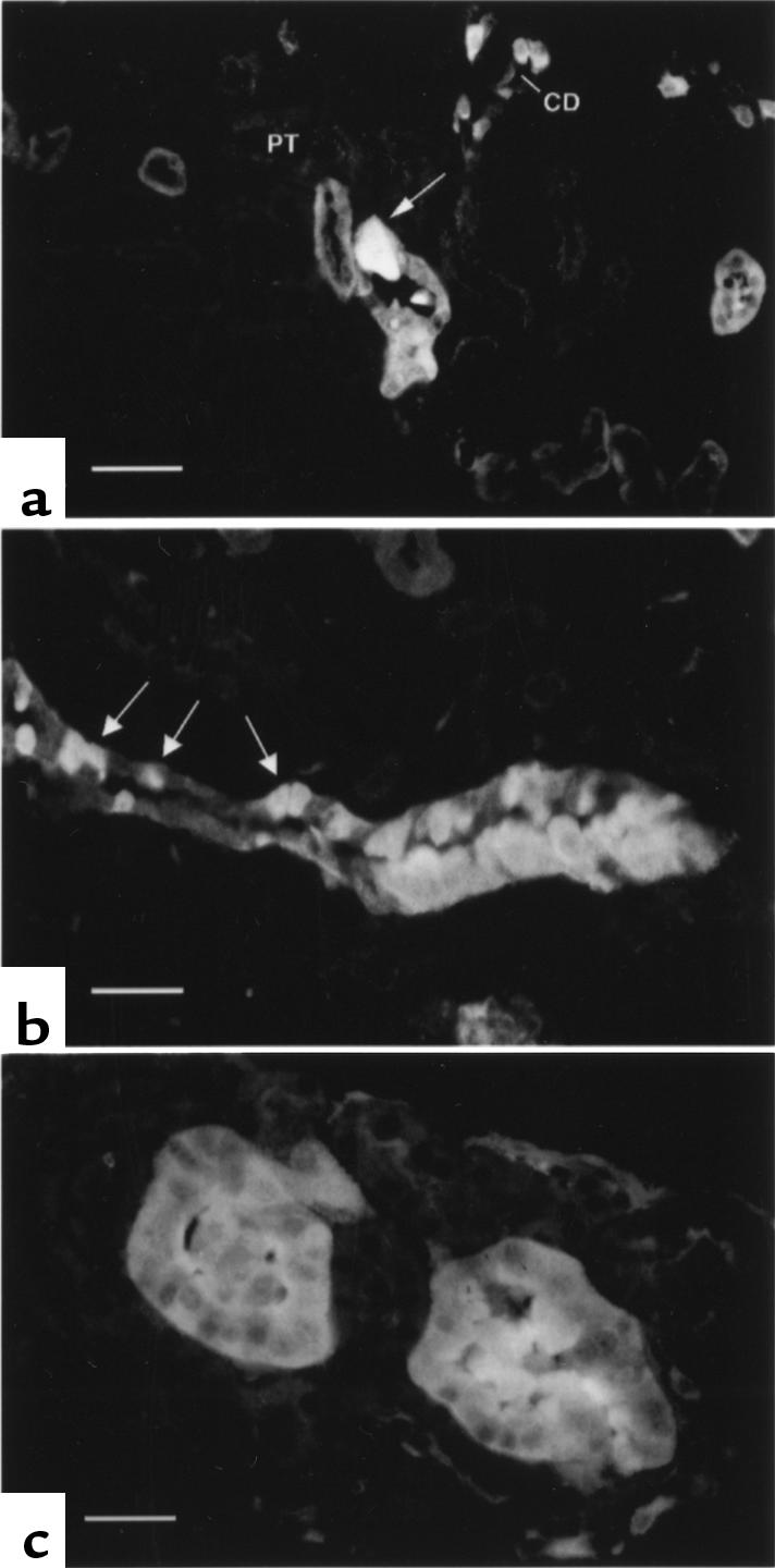
Detection of early renal neoplastic lesions using gelsolin immunolocalization. (a) A single atypical cortical collecting duct cell with intense gelsolin staining in an otherwise normal cortical collecting duct. Proximal tubules (PT) and cortical collecting ducts (CD) are indicated. (b) A cluster of atypical cells highly expressing gelsolin are seen in a cortical collecting duct that also contains normal tubular cells. Normal intercalated cells are indicated by arrows. (c) An early-stage solid adenoma shows high-level gelsolin expression in all cells. Scale bars are 25 μm (a and b) and 15 μm (c).