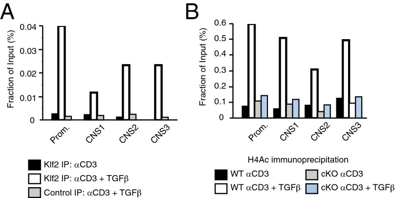
Fig. 4.
KLF2 directly promotes FoxP3 transcription within the iTreg lineage. (A) ChIP assays using antibody directed against KLF2 after lysis of wild-type CD4+CD25− T cells cultured under conditions that promote T-cell activation (black) vs. iTreg differentiation (white). Isotype control antibody (gray) is included for iTreg ChIP assays. DNA primers are specific for the promoter or enhancer elements (CNS1-3) of FoxP3. KLF2 binding to the regulatory regions of FoxP3 is graphed relative to input amounts of genomic DNA. These experiments were performed three times. (B) Wild-type (WT) and KLF2-deficient (cKO) CD4+CD25− T cells were cultured under activating (black, gray) vs. iTreg-inducing conditions (white, blue) before performing ChIP assays using antibody directed against acetylated histone H4. This experiment was conducted twice.

