
Fig. 3.
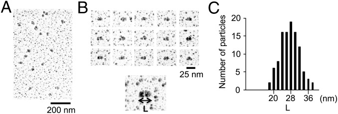
Structural analysis of DC1–2–3. (A) Low-magnification electron microscopy images of DC1–2–3 prepared by low-angle rotary shadowing. (B) Typical images of DC1–2–3. Each particle has an ellipsoidal shape. (C) Histogram of the major axis length of DC1–2–3 (L) (n = 108). Considering the thickness of platinum coating (2 nm on all sides), the peak length is ∼24 nm.