Fig. 5.
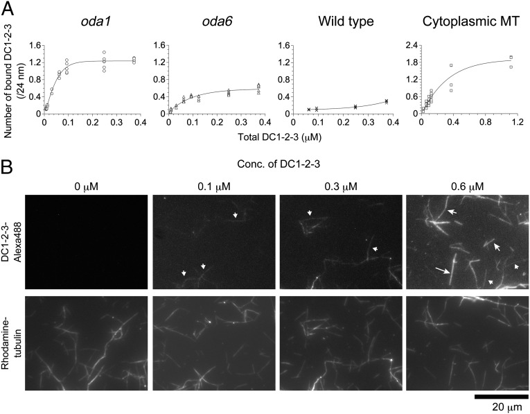
Cooperative binding of DC1–2–3 to microtubules. (A) Binding between DC1–2–3 and axonemes or cytoplasmic microtubules was analyzed by cosedimentation assay. Purified DC1–2–3 was mixed with axonemes of oda1, oda6, and wild-type cells, and with porcine brain cytoplasmic microtubules. The amount of bound DC1–2–3 was calculated as the number of molecules bound to microtubules per 24 nm (y axis), and plotted against total DC1–2–3 (x axis). The saturated amount of bound DC1–2–3 was in the order of cytoplasmic microtubules > oda1 > oda6 > wild type, reflecting the available sites for ODA-DC binding. (B) Different concentrations of Alexa-488–labeled DC1–2–3 were mixed with rhodamine-labeled microtubules and observed by fluorescence microscopy. At 0.1 µM DC1–2–3, some microtubules were labeled whereas others were not at all. At 0.3 µM, more microtubules were labeled but some remained unlabeled. At 0.6 µM, almost all microtubules were labeled. (B, Upper) The lengths of the arrows represent the fluorescence intensity, and microtubules marked with the same size of arrows have similar intensities. Shortest arrows are ∼30 a.u., mid-size arrows ∼60 to ∼70 a.u., and the longest arrow ∼87 a.u.

