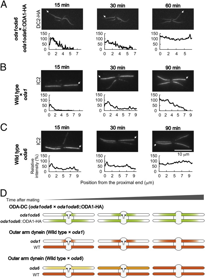
Fig. 6.
The ordered binding of the OAD to the axoneme is dependent on the ODA-DC. (A–C, Upper) Immunofluorescence staining of temporary dikaryons: (A) oda1oda6×oda1oda6::ODA1–HA expressing DC2–3×HA, (B) wild type×oda1, and (C) wild type×oda6. The time after mating is noted above each panel. Dikaryons were demembranated and labeled with (A) anti-HA or (B and C) anti-IC2 antibody. (A–C, Lower) The fluorescence intensity of DC2-HA or IC2 in axonemes originally lacking those proteins was normalized respectively with that of oda1oda6::ODA1–HA (A, arrows) or wild-type (B and C, arrows) axonemes in the same dikaryon. (A) Binding of the HA-tagged ODA-DC proceeded from the proximal part of the oda1oda6 axoneme to the distal part. (B) Similarly, binding of OAD proceeded from proximal to distal in the oda1 axoneme. (C) In contrast, binding of OAD gradually increased over the whole length of the oda6 axoneme. The difference is that the ODA-DC was already present on the doublets of the oda6 axonemes but not on the doublets of the oda1 axonemes; hence, ODA assembly was constrained to follow the pattern of ODA-DC assembly in the oda1 axonemes. (D) Schematic diagrams illustrating patterns of labeling for the ODA-DC (green) and the OAD (shades of orange) in the experiments of A–C.

