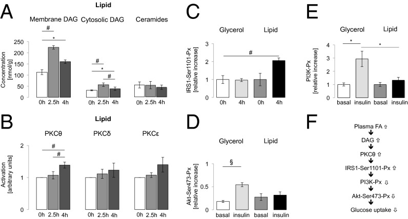
Fig. 1.
(A) Myocellular DAG concentrations in the membrane and cytosolic fraction and myocellular ceramide concentrations during lipid infusion in young lean healthy controls (CON) (n = 10). (B) Activation of myocellular PKCθ, -δ, and -ε during lipid infusion in CON (n = 10–14). (C) Phosphorylation of serine 1101 residue at IRS1-Ser1101-Px at baseline and its relative increase after 4 h glycerol (white and light gray columns) or lipid (dark gray and black columns) infusion in young lean healthy controls (CON) (n = 7). (D and E) PI3K-Px (E) and membrane/cytosolic ratio of Akt-Ser473 phosphorylation (Akt-Ser473-Px) (D) at baseline and after 4.5 h of glycerol (light gray column) or lipid (black column) infusion during insulin stimulation for 30 min in CON (n = 7). Data are given as means ± SEM. *P < 0.05; #P < 0.01; §P < 0.001. (F) Increased plasma FAs lead to myocellular accumulation of DAGs and consequent IRS1-Ser1101-Px, impaired PI3K-Px, and blunted insulin stimulation of Akt-Ser473-Px.

