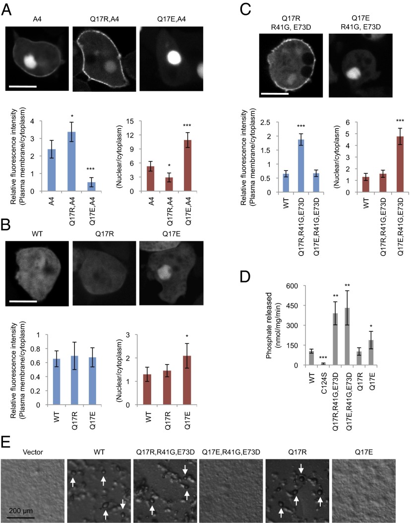
Fig. 2.
Q17R promotes PTEN membrane association in PTENA4 and PTENR41,E73D. (A–C) Dictyostelium cells expressing the indicated forms of PTEN-GFP were observed by fluorescence microscopy. (Scale bar, 10 µm.) Intensity of GFP at the plasma membrane was quantified relative to that in the cytosol. Values represent the mean ± SD (n ≥ 15). (D) PTEN-GFP, PTENC124S-GFP, PTENQ17R-GFP, and PTENQ17E-GFP were immunopurified from Dictyostelium cells, and phosphatase activities were measured (n ≥ 3). (E) PTEN-null Dictyostelium cells expressing different PTEN-GFP constructs were starved for 36 h to induce differentiation into fruiting bodies (white arrows).

