Fig. 5.
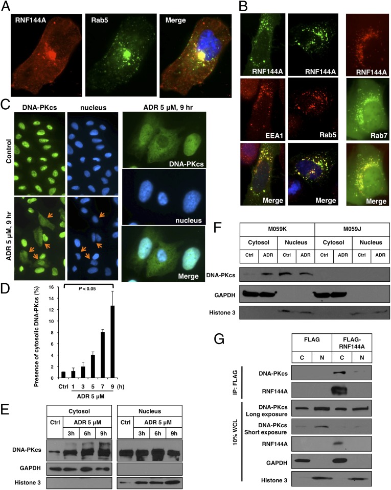
RNF144A localizes at endosomes and interacts with cytoplasmic DNA-PKcs. (A and B) RNF144A localizes at endosomal organelles. (A) The immunofluorescence images (100×) show that RNF144A (red) partially overlapped with Rab5, an endosomal marker, and (green) positive dots. FLAG-tagged RNF144A was transfected into U2OS cells. On the next day, cells were fixed and stained with anti-FLAG antibody (red) and anti-Rab5 antibody (green). (B, Left and Center) EGFP-tagged RNF144A was transfected into U2OS cells for 1 d. Cells were fixed and stained with EEA1 or Rab5 antibody and a Texas Red-conjugated secondary antibody. (B, Right) mCherry-tagged RNF144A was cotransfected with EGFP-tagged Rab7 (48) into U2OS cells for 1 d. Immunofluorescence images (100×) were acquired by a Zeiss inverted microscope. Expanded image data are shown in Fig. S4. (C–F) ADR-induced redistribution of DNA-PKcs from the nucleus to the cytoplasm. U2OS cells were treated with ADR (5 μM) for the indicated time, and then, they were fixed and stained with anti–DNA-PKcs (green) and Hoechst 33258 (blue). (C) Orange arrow indicates the cells with cytoplasmic DNA-PKcs staining. High-magnification images of the ADR-treated cells are shown in Right. The specificity of anti–DNA-PKcs antibody for immunofluorescence study was verified using a pair of glioma cells: M059K (DNA-PKcs–proficient) and M059J (DNA-PKcs–deficient) cells (Fig. S6). (D) The percentages of cells with cytoplasmic DNA-PKcs staining on ADR treatment were counted (from 1,000 cells) and shown. Error bars represent SDs from three independent experiments. (E) Western blot analysis shows ADR-induced cytoplasmic DNA-PKcs in a time-dependent manner. (F) M059K (DNA-PKcs–proficient) and M059J (DNA-PKcs–deficient) cells were treated with ADR (5 μM) for 9 h, and then, they were harvested for fractionation and Western blot analysis. (G) FLAG-tagged RNF144A interacted with cytoplasmic DNA-PKcs. An empty FLAG vector or FLAG-tagged RNF144A was transfected to HEK293T cells. After 24 h, cell fractionation was performed. Cytosolic (C) or nuclear (N) extracts were then immunoprecipitated by FLAG M2 beads and immunoblotted with indicated antibodies. Ctrl, control.

