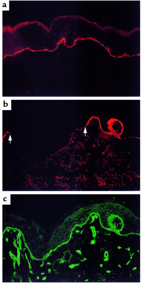
Figure 1.
Type XVII collagen was present only at focal sites in the proband’s epidermal BM. (a) IF microscopy studies of normal human skin cryosections stained with an mAb to type XVII collagen (see Methods) showed continuous expression of this protein in epidermal BM (shown in red). (b) IF microscopy studies of the proband’s skin cryosections stained in the same manner found that type XVII collagen (when detected) was present in epidermal BM only in a focal, interrupted distribution (shown in red; an intervening segment of nonreactive BM is delineated by arrows). (c) The proband’s epidermal BM was otherwise intact. For purposes of documentation, the cryosection shown in b was double stained with an antibody to type IV collagen. This antibody (as well as antibodies to bullous pemphigoid antigen 1, laminin 5, and type VII collagen; ref. 9) showed normal reactivity (shown in green) along the entire epidermal BM of the proband. Type IV collagen in microvascular BMs was also stained.

