Figure 2.
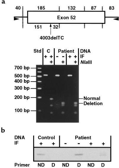
A normal COL17A1 allele was not present in the proband. (a) LCM was used to collect epidermal cells overlying segments of the proband’s epidermal BM that stained positive (+) or negative (–) for type XVII collagen by IF microscopy (IF); epidermis collected from normal human skin by LCM served as a control (C). Genomic DNA was isolated from these samples, and exon 52 of COL17A1 was PCR amplified and analyzed by restriction endonuclease digestion. For illustration, the schematic depicts exon 52 (rectangle) and its flanking intronic regions (solid lines) that were amplified by PCR (primers indicated by arrowheads). Vertical dotted lines signify NlaIII restriction endonuclease sites in PCR products derived from a normal COL17A1 allele; the sizes of the resulting fragments (in base pairs) are listed along the top of the schematic. The arrow directed to the underside of exon 52 shows the NlaIII cleavage site introduced by 4003delTC; this additional NlaIII restriction site cleaves the 185-bp fragment into segments of 151 and 32 bp as indicated. The PCR products outlined above, and a 100-bp ladder standard (Std), were electrophoresed on a 3% agarose gel and then stained with ethidium bromide. Undigested PCR products derived from control DNA, as well as negative-patient and positive-patient DNA, all comigrated as shown. NlaIII restriction analysis of these PCR products revealed that control DNA was derived from normal COL17A1 alleles and that both negative-patient and positive-patient DNA were derived from alleles bearing 4003delTC. Note that the 32- and 40-bp fragments are not apparent in this gel. (b) That positive-patient DNA was not derived from normal COL17A1 alleles was confirmed by allele-specific PCR using primers ending on sequences specific to the normal (ND, no deletion) or mutant (D, deletion) alleles at nucleotide 4003. Analysis of these PCR products on 4–20% polyacrylamide gels confirmed the normal sequence in control DNA and showed that both negative-patient and positive-patient DNA were amplified from alleles containing 4003delTC. The presence of a doublet in the positive-patient (but not the negative-patient) DNA using the D primers suggested heterozygosity for a mutation downstream from 4003delTC.

