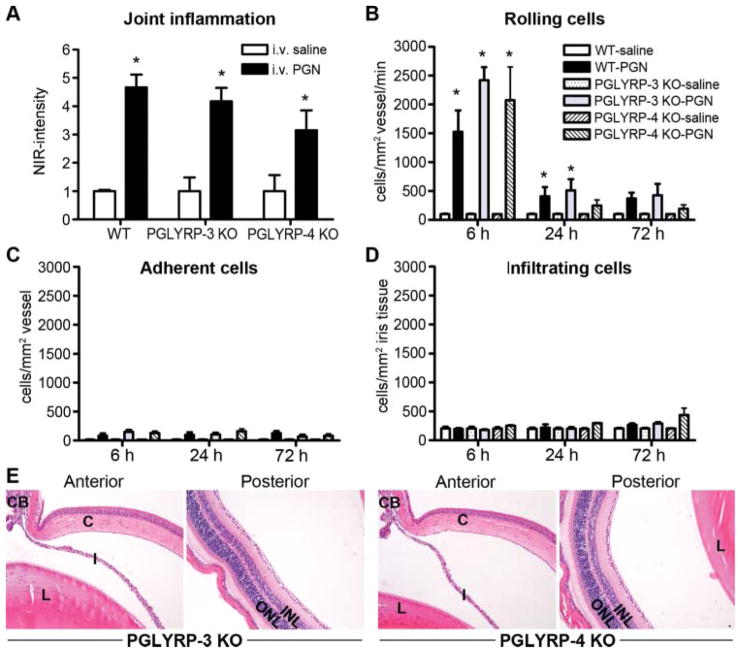
Figure 4.
Assessment for a contribution of PGLYRP-3 or PGLYRP-4 in arthritis and uveitis triggered by systemic peptidoglycan (PGN) exposure. Mice were systemically administered PGN or saline. Severity of ankle inflammation was assessed by near-infrared-imaging at 72 h postinjection (A). The leukocyte trafficking response within the iris was documented by intravital microscopy, and the number of rolling (B), adherent (C) and infiltrating (D) cells was quantified. At 72 h following PGN exposure, uveitis was assessed histologically and panel E depicts representative histological images of anterior and posterior eye segments of PGLYRP-3 and PGLYRP-4 knockout mice (H&E stain, original magnification at 200 ×). *p<0.05 comparison to saline controls within a genotype (n=10 mice/treatment/genotype). H&E, haematoxylin and eosin.

