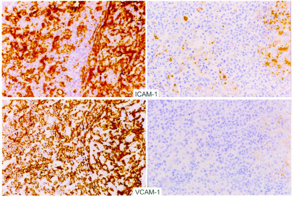
Figure 6.
Representative photomicrographs demonstrating immunohistochemical staining of lymph node sections before (left) and after (right) HAART with ICAM-1 (top) and VCAM-1 (bottom). Expression of these markers was decreased in each pair of biopsies examined. These sections represent analysis performed on the same day with the same staining conditions for biopsies obtained from an individual patient.

