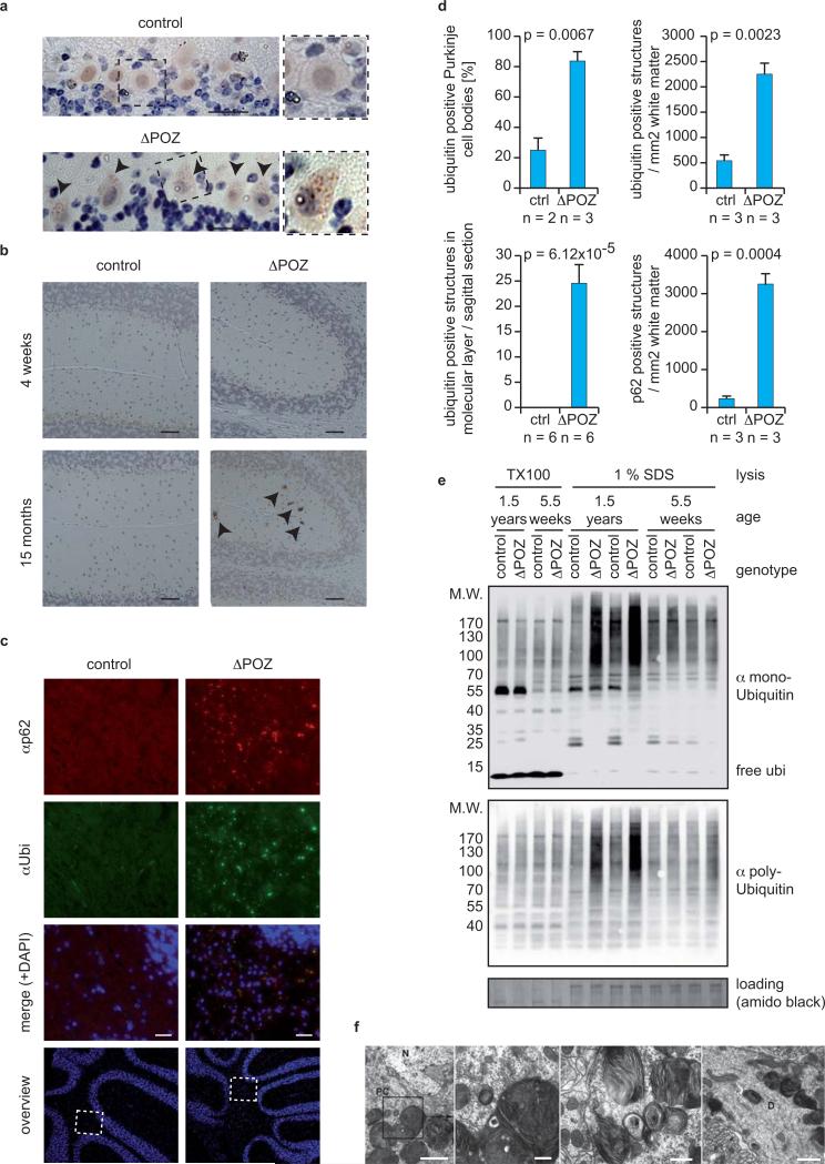
Figure 5. Ubiquitinated proteins and p62/Sqstm1 accumulate in an age-dependent manner in cerebella of Miz1ΔPOZNes mice.
(a,b) Immunostaining using an α-ubiquitin antibody of (a) Purkinje cells and (b) the molecular layer of Miz1ΔPOZNes and age-matched control mice (dotted area: higher magnification). Arrowheads designate ubiquitin-positive structures. Scale bar: (a) 25 μm; (b) 50 μm.
(c) Immunofluorescence staining using an α-p62 and an α-ubiquitin antibody of the white matter of cerebella from Miz1ΔPOZNes and age-matched control mice. Nuclei were counterstained with DAPI and overviews of the cerebella clarifying the selected areas are shown. Scale bar: 25 μm.
(d) Quantification of a, b and c. Error bars represent SEM derived from the indicated numbers of animals. p-values were calculated using an unpaired two-tailed Student's t-test.
(e) Immunoblots of Triton X-100 soluble (“TX100”) and insoluble (“1% SDS”) fractions of cerebella obtained from control and Miz1ΔPOZNes mice of the indicated ages. Blots were probed with antibodies recognizing either mono- or polyubiquitinated proteins. Staining of the membrane with amido black was used as loading control.
(f) Transmission electron microscopy documenting the accumulation of large multilamellar bodies in Purkinje cell bodies (PC) and Purkinje dendrites (D) of Miz1ΔPOZNes mice (N: nucleus). Such structures were not observed in age-matched control mice (as quantified in Supplementary Figure S7b). Mean age of mice for all electron microscopy pictures is 13 months for Miz1ΔPOZNes and 12.5 months for control mice. Morphologically similar structures accumulate in neurons of mice deficient in late steps of autophagy 28. Scale bars: 2, 0.2, 0.5 and 1 μm, respectively.

