
Figure 4.
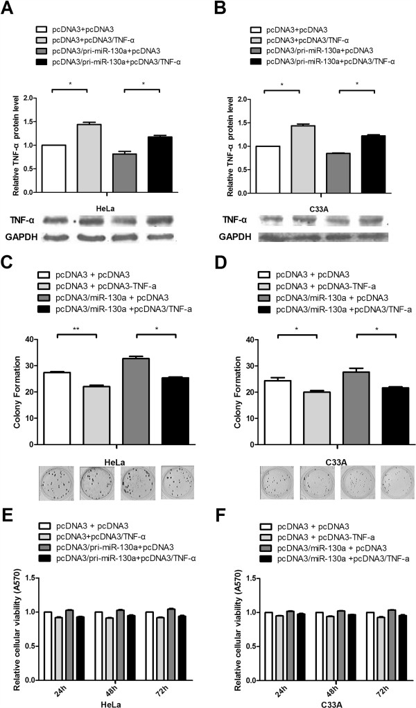
TNF-α overexpression counteracts the effects of miR-130a. (A and B) Western blot analysis was used to determine TNF-α protein levels and validate the TNF-α overexpression vector. HeLa and C33A cells were co-transfected with the pcDNA3/TNF-α plasmid, which did not contain the TNF-α 3’UTR, with or without the pcDNA3/pri-miR-130a plasmid. GAPDH protein expression was used to normalize the endogenous expression data. (C and D) Colony formation assays were performed to determine the effects of TNF-α on cell proliferation. We transfected the pcDNA3/TNF-α vector with or without cotransfection of pcDNA3/pri-miR-130a as in Figure 2G and H. (E and F) MTT assay used to assess cell viability. The data represent the mean ± SD of three independent experiments. (*P < 0.05, **P < 0.01).