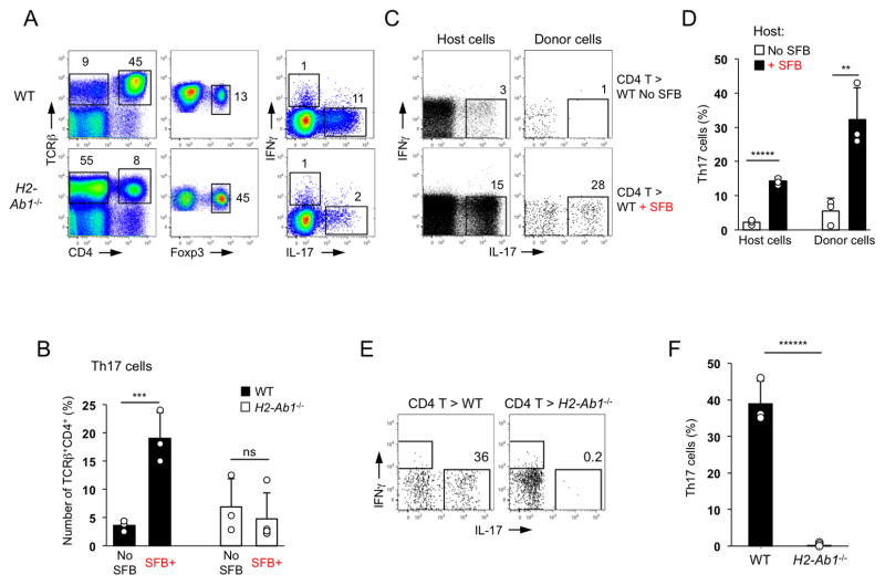
Figure 1. Induction of intestinal Th17 cells by SFB requires MHCII expression in the periphery.
A. Th17 and Treg cell proportions in SI LP of SFB-positive WT and MHCII-deficient (IAb−/−) mice. Foxp3 and cytokine staining plots are gated on TCRβ+CD4+ cells
B. Th17 and Treg cell proportions in SI LP of SFB-negative (Jackson microbiota) and SFB-positive (Taconic microbiota) WT and IAb−/− mice. Plots gated on TCRβ+CD4+ cells
C–D. WT CD45.1+ CD4 T cells were adoptively transferred into WT CD45.2 mice before or 12 days after SFB colonization. Cytokine expression in host (CD45.2+) and donor (CD45.1+) SI LP TCRβ+CD4+ cells 2 weeks after transfer. Data from one of multiple experiments
E–F. Th17 cell induction in WT CD4 T cells two weeks after transfer into SFB-positive WT and MHCII-deficient recipients. Plots gated on TCRβ+CD4+ cells. Data from one of multiple experiments

