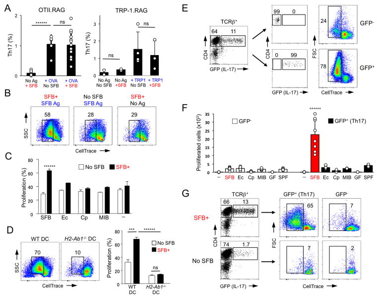
Figure 2. SFB-induced intestinal Th17 cells preferentially respond to SFB antigens.
A. Th17 cell proportions in the SI LP of OTII.RAG and TRP-1.RAG TCR Tg mice before and after SFB colonization in the absence or presence of cognate antigen. Representative data from 5 independent experiments
B–C. Proliferation response of sorted SI LP TCRβ+CD4+ cells from SFB-negative (Jax) and SFB-positive (Tac) WT B6 mice to SFB (B, C) or other bacterial antigens (C). T cell proliferation was scored by dye dilution on Day 3. Ec, E. coli, Cp, Clostridium perfringens; MIB, mouse intestinal bacteria (cultured isolates from feces of SFB-negative (Jackson) mice); “-” – no antigen. Representative data from 5 independent experiments
D. SI LP TCRβ+CD4+ cells were purified from SFB-negative (No SFB) and SFB-positive (SFB+) WT mice and co-cultured with SFB antigens as in (B) and WT or IAb−/− DCs. Data from 2 independent experiments
E–F. SI LP GFP+ (Th17) and GFP− (non-Th17) TCRβ+CD4+ cells from SFB-positive Il17GFP mice were stimulated in vitro with SFB (E, F) or various bacterial antigens (F) as in (B) or with lysates from germ-free (GF) or SFB-negative SPF (SPF) animals. Representative data from multiple experiments
G. SI LP GFP+ (Th17) and GFP− (non-Th17) TCRβ+CD4+ cells from SFB-positive (SFB+) or SFB-negative (No SFB) Il17GFP mice were stimulated in vitro with SFB antigens as in (B). Representative data from 2 independent experiments

