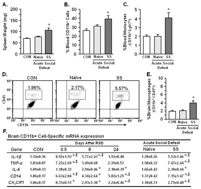
Figure 4.
Acute social defeat enhanced the neuroinflammatory profile and reinitiated macrophage recruitment to the brain of stress-sensitized mice. These studies included three experimental groups of mice: 1) control (CON) nonstressed mice that were not exposed to any social defeat; 2) naïve (no initial stress) mice subjected to acute social defeat 24 days later; and 3) stress-sensitized (SS) mice that were exposed to repeated social defeat (RSD) and subjected to acute social defeat 24 days later. Following behavioral testing (Figure 3), spleen, blood, and brain CD11b+ cells were collected. Acute social defeat markedly increased (A) spleen weight (F2,36 = 12.51, p < .0001), (B) blood myeloid (CD11b+) cells (F2,35 = 6.01, p < .006), and (C) percentage of blood monocytes (CD11b+/Ly6Chi) in stress-sensitized mice (F2,29 = 7.23, p < .003). (D) Representative flow bivariate dot plots of CD11b/CD45 labeling in Percoll isolated microglia/macrophages show that acute social defeat also increased (E) the percentage of brain macrophages (CD11b+/CD45hi) in SS mice (F2,26 = 10.72, p < .0005). Bars represent the mean ± SEM. (F) Corresponding with increased macrophage recruitment in the brain, RSD increased relative gene expression of interleukin-1 beta (IL-1β), tumor necrosis factor-alpha (TNF-α), interleukin-6 (IL-6), and CD14, (p < .03 for each) and decreased CX3CR1 (p < .05) in enriched brain CD11b+ cells. In addition, acute social defeat enhanced expression of all genes analyzed in enriched brain CD11b+ cells from stress-sensitized mice (p < .05 for each). Values represent average fold change ± SEM. Means with asterisk (*) are significantly different from CON (p < .05) and means with (‡) are significantly different from naïve (p < .05). mRNA, messenger RNA.

