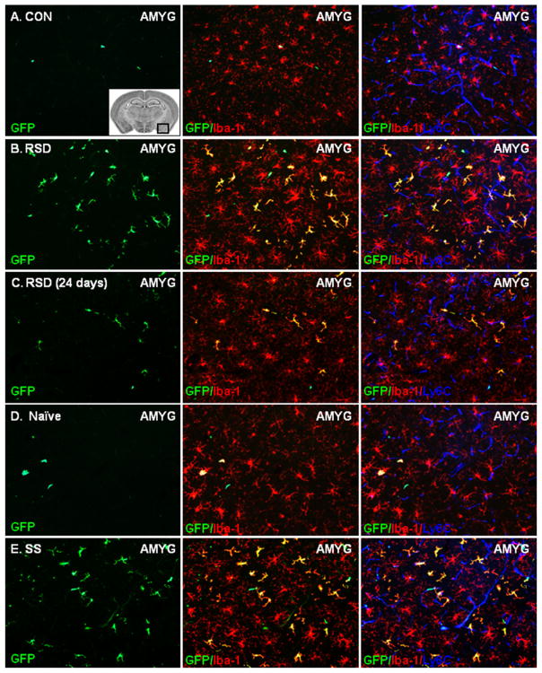
Figure 6.
Acute social defeat promoted infiltration of bone marrow (BM)-derived macrophages into the brain of stress-sensitized (SS) mice. Brains were collected from control (CON), repeated social defeat (RSD) (.5, 8, or 24 days later), naïve, and stress-sensitized green fluorescent protein (GFP)+ BM-chimera mice. Representative images of peripheral GFP+ (green) cells in the amygdala (AMYG) of (A) CON, (B) RSD (.5 days), (C) RSD (24 days), (D) naïve, and (E) SS mice are shown. The inset image illustrates the area of the AMYG in which the representative images were taken (64). Sections were co-labeled with antibodies to identify resident microglia (ionized calcium binding adaptor molecule 1 [Iba-1]+, red), the vasculature (Ly6C+, blue), and parenchymal BM-macrophages (GFP+/Iba-1+, yellow). The subsequent panels in each row show merged GFP/Iba-1 or GFP/Iba-1/Ly6C images. White scale bar represents 100 μm (20×).

