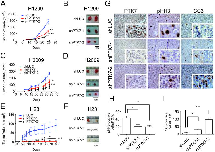
Figure 6.

PTK7 depletion in three NSCLC cell lines decreases tumor burden in a xenotransplantation assay. A. Tumor volume over time of xenografted H1299 with control shRNA against luciferase (shLUC), or 2 independent hairpins against PTK7. Error bars represent standard error of the mean (s.e.m) and significant p-values correspond to <0.01 (**) or <0.001 (***) when compared to the shLUC control. Values are mean ± SEM (n = 4-8). B. Representative ex vivo images of H1299 tumors. C and D. Same figures for H2009 cell line. E and F. Same figures for H23 cell line. G. Staining of PTK7, pHH3 and CC3 by IHC in the H1299 xenografts. H. Quantification of pHH3-positive cells per field of view (n=6) I. Quantification of CC3-positive cells per field of view (n=6).
