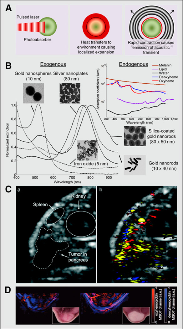
FIGURE 2. Photoacoustic molecular imaging of cancer.
(A) Diagram of photoacoustic effect. Pulsed laser irradiation is absorbed by photoabsorber, causing localized heating and expansion of directly surrounding environment. During rapid contraction, high-frequency acoustic transient (sound wave) is emitted. (B) Absorption spectra of endogenous and exogenous contrast agents. (C) Ultrasound (left) and spectroscopically resolved photoacoustic images (right) (14.5 × 11.8 mm) of epidermal growth factor receptor targeted silver nanoplates (yellow), oxygenated hemoglobin (red), and deoxygenated hemoglobin (blue) in human pancreatic carcinoma (MPanc96 cells) tumor xenograft (21). (D) Spectroscopically resolved photoacoustic images (and corresponding photographs) of oxy- and deoxyhemoglobin in orthotopic murine breast tumor in mice (20). a.u. = arbitrary unit; MSOT = multispectral optoacoustic tomography. (Silicacoated gold nanorods and iron oxide nanoparticle images and spectra in B reproduced with permission of (24,25); C reproduced with permission of (21); D reproduced with permission of (20).)

