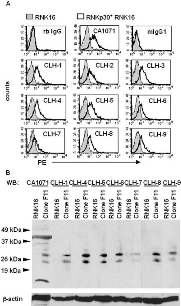
Fig. 1.
Anti-rNKp30 mAb specifically detect rNKp30 on transfected cells. (A) Anti-rNKp30 pAb, CA1071, anti-rNKp30 mAb CLH-1 through CLH-9, and isotype control antibodies followed by PE-conjugated secondary antibodies were used to stain parental RNK16 cells (filled histograms) and stably transfected rNKp30+ RNK16 Clone F11 cells (open histograms). (B) Immunoblot analysis of lysates from parental RNK16 and rNKp30+ Clone F11 cells were performed and blots were probed with anti-rNKp30 antibodies as specified and reprobed for β-actin. Molecular masses (kDa) of a protein size ladder are indicated on the left by arrowheads. rNKp30 antibodies detect two prominent bands between 25 and 30 kDa specifically in the rNKp30+ RNK16 clone F11 lysates (n=3).

