Fig. 3.
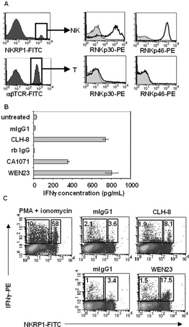
rNKp30 is expressed on a major subset of F344 spleen NK cells and activation induces IFN-γ production. (A) rNKp30 and rNKp46 expression were analyzed on F344 rat spleen NK cells (NKRPl-FITChi, αβTCR-PerCP−) and T cells (αβTCR-FITC+) by flow cytometry. NKp30 expression was detected using CLH-9-biotin (middle, open histograms) and NKp46 expression was identified with WEN23-biotin (right, open histograms). NCR histograms were overlayed on isotype controls (filled histograms). (B) Splenocytes or PBMC (2.5 × 10⁶/mL, 5 × 10⁵ cells/ well) were cultured in media alone (untreated), or with immobilized antibodies (12 µg/mL). Splenocytes were treated with antibodies against rNKp30 (CLH-8 or CA1071), or control antibodies (mIgG1 or rb IgG) for 11 h. As a control for NK-mediated IFN-γ secretion, PBMC were treated with the anti-rNKp46 mAb, WEN23. Harvested supernatants were assessed for IFN-γ production by ELISA (n=5). (C) Treated mononuclear cells were permeabilized and analyzed for IFN-γ with a PE-conjugated anti-IFN-γ mAb, and costained with a FITC-conjugated anti-NKRPl antibody (n=3). Each dotplot contains a small box (right) indicating the percent of IFN-γ+ NK cells, and a large box (left) gating on the fraction of non-NK cells expressing IFN-γ. (Top row) Splenocytes were treated with PMA (10 ng/mL) and ionomycin (0.5 µg/mL) (left), mIgG1 isotype control (12 µg/mL) (middle), and anti-rNKp30 mAb CLH-8 (12 µg/mL) (right). (Bottom row) PBMC were treated with mIgG1 isotype control antibodies (12 µg/mL) or anti-rNKp46 mAb WEN23 (12 µg/mL). rNKp30 and rNKp46 activation induced increased IFN-γ production from a subset of NK cells.

