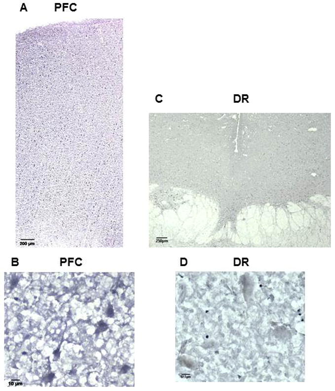
Fig. 3.

Distribution of Freud-2 immunoreactivity in human prefrontal cortex and dorsal raphe nuclei. Frozen sections of post-mortem human prefrontal (PFC; sections A and B) and dorsal raphe nuclei (DR; sections C and D) were incubated with anti-Freud-2 antibody and processed for immunohistochemistry using anti-Freud-2; no specific staining was observed using preimmune serum (not shown). Sections B and D are at high magnification (40x). Freud-2 staining is enriched in grey matter of prefrontal cortex, but sparse in dorsal raphe nuclei (DR).
