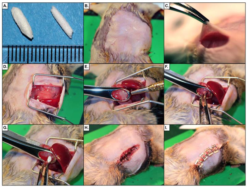
Figure 3. Demonstration of Abdominal Laparotomy Procedure.
(A) Rolled Gelfoam® plugs are shown with a mm scale. Plugs are used to fill the biopsy wound and encourage clotting. (B) The surgical site is prepared by removing fur and disinfection with surgical scrub. (C) A 15mm incision is made in the skin, followed by a 12mm incision in the abdominal wall. (D) Retractors are inserted into the abdomen to create an operative field. (E) The tumor is isolated and clamped in the biopsy forceps, taking care not to rupture an nearby vessels. The biopsy punch tool is then passed through the tumor to the back plate of the biopsy forceps to cut a cylinder of tumor tissue. (F) The biopsy core is removed with a pair of forceps. (G) A Gelfoam® plug is inserted into the wound left from the biopsy. (H) The abdominal wall is sutured with 5-0 silk. (I) The skin incision is repaired with surgical staples.

