
Figure 1.
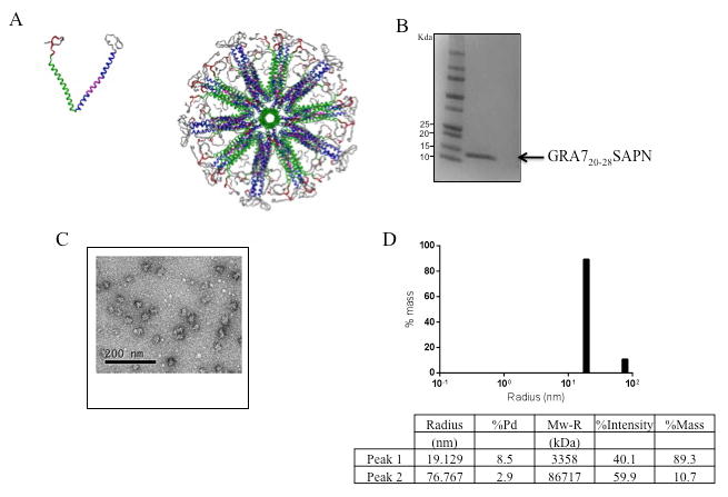
Assembly of GRA720-28 SAPN. A, Three-dimensional model of the nanoparticle. Left: single protein chain of the nanoparticle. Right: assembled nanoparticle with icosahedral symmetry viewing down the five-fold symmetry axis. Green: pentameric coiled coil; Blue: trimeric coiled coil; Magenta: CD4 epitope as an integral portion of the trimeric coiled coil; red: CD8 epitope; Gray: His-tag; Light gray: solubilization tag. B, SDS-PAGE 4-20% of the purified LP. C, Transmission electron microscopy of a nanoparticle preparation containing one CD8+ T cell epitope from dense granule protein GRA7 (GRA720–28) and the universal CD4+ TH epitopte (PADRE). D, DLS plot of GRA720-28 nanoparticles after final purification and assembly. For this product the diameter of 89.3% of the protein is ∼38nm.