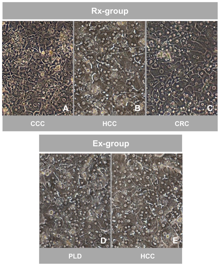
Figure 2. Comparable morphological appearance of cultured hepatocytes isolated from resected surgical specimens and explanted diseased livers.
Phase-contrast microscopy of cultured (day 5) primary human hepatocytes isolated from resected surgical specimens (Rx-group) removed due to metastasis of colorectal cancer (CRC) (A), hepatocellular carcinoma (HCC) (B) and cholangiocellular carcinoma (CCC) (C) as well as from explanted diseased livers (Ex-group) due to HCC (D) and polycystic liver disease (PLD) (E). Hepatocytes of both groups show the typical polygonal shape, are highly prismatic and either mono- or polynucleated. Signs of the formation of bile canaliculi are present. Magnification 100x.

