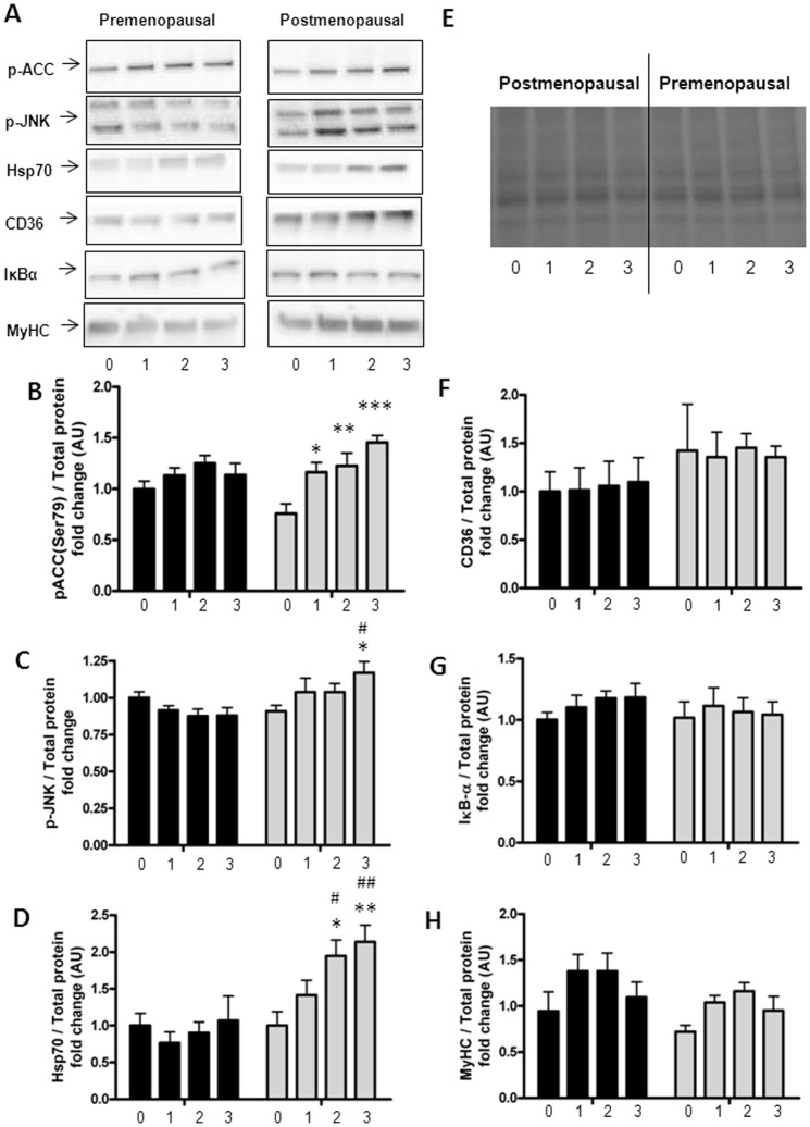
Figure 2. Protein expression in palmitate treated myotubes from premenopausal and postmenopausal women.
Satellite cells were isolated from vastus lateralis biopsies from premenopausal and postmenopausal women. Cells were grown in culture until mature myotubes were formed. Myotubes were treated with palmitate in the last one, two or three days of differentiation. Protein lysates from the myotubes were immunoblottet to assess the protein expression of specific proteins as stated below. Protein expression in pre-myotubes (n = 6, black bars) and post-myotubes (n = 5, white bars) in response to palmitate treatment, (A) Representative immunoblots of all investigated proteins, (B) p-ACC-Ser79 (280 kDa) was increased only in post-myotubes in response to palmitate treatment, (C) p-JNK-Thr183/Tyr185 (54/46 kDa) was increased after three days of palmitate treatment in post-myotubes and reached significantly higher levels than in post-myotubes, (D) Hsp70 (72 kDa) was increased after two and three days of palmitate treatment in post-myotubes, and reached significantly higher levels than in pre-myotubes, (E) Representative blot of total protein, (F) CD36 (88 kDa) was unaffected by both palmitate treatment and menopausal status, (G) IκBα (39 kDa) was unaffected by both palmitate treatment and menopausal status, (H) MyHC (230 kDa) was unaffected by both palmitate treatment and menopausal status. Significantly different from control; * p<0.05, ** p<0.01, *** p<0.001. Significantly different from premenopausal, at same palmitate exposure time; # p<0.05, ## p<0.01. 0: Zero days of palmitate treatment (control), 1: One day of palmitate treatment, 2: Two days of palmitate treatment, 3: Three days of palmitate treatment. All data were normalized to the premenopausal control group. Data are presented as means ± SEM.

