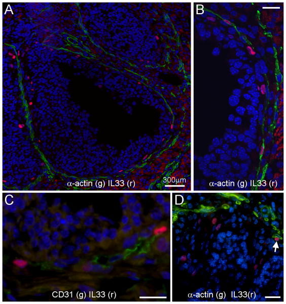
Figure 2. Tissue location of ovarian cells with nuclear IL33 (nIL33+ cells) in mice during hCG-induced ovulation as detected by immunofluorescence.
A. An ovulating mature follicle shows many nIL33+ cells (red) around inner layer of its theca. Note that nIL33 staining (red) is colocalized to the cells, which express smooth muscle α-actin (SMα-actin, green). B. An enlarged view of co-localization of nIL33+ (red) with SMα-actin+ (green) cells. C. nIL33+ cells (red) in the theca of ovulating follicles also express CD31 (green). D. A group of nIL33+ cells (red) are mingled with granulosa cells of an ovulating follicle with a distance from SMα-actin+ cells (green). Note different nuclear morphology of granulosa cells and nIL33+ cells. A SMα-actin associated nIL33+ cell is also shown (arrow). Nuclei were counter stained by DAPI. Bars in B, C and D = 20 μm.

