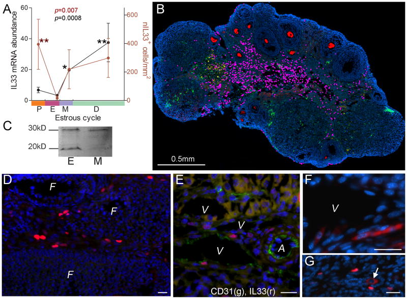
Figure 4. IL33 expression during estrous cycle in mice.
A. Summary of quantitative PCR detection of IL33 mRNA (black dots and line, left y-axis) and numbers of nIL33+ cells (red dots and line, right y-axis) during estrous cycle. Estrous stage in an individual was determined by vaginal smear; theoretical duration for each stage follows a published paper (32). P, proestrus; E, estrus, M, metestrus; D, diestrus. Cell numbers are expressed as cell density (cell number/mm2). Kruskal-Wallis test was applied; data for each group was compared to that for estrus with Dunn’s post test. B. A representative merged super-image covering a whole ovarian section at diestrus after three-color immunofluorescence is shown. Each nIL33+ cell is labeled by a pink dot for identification. Cytoplasm of oocytes is stained red. Nuclei were counter-stained by DAPI. C. Western blot detection of two forms of IL33 (30kD for nuclear form and 20kD for cleaved form) in the ovaries at estrus (E) and metestrus (M). Note a higher level of cytokine form of IL33 at estrus as compared to the lowest number of nIL33+ cells during estrus cycle (A) at this stage. D. Immunofluorescence shows a group of nIL33+ cells (red) in the veins which surround various follicles (F). E. Two color immunofluorescence shows nIL33+ cells (red) to be CD31+ endothelial cells (green); V, vein; A, artery. F. A group of cells with cytoplasmic IL33 (red) are observed in endothelia of a vein (V) at estrus stage. G. A cell with cytoplasmic IL33 (arrow, red) is shown to be among many nIL33+ cells (red) at other stages. Bars in D, E, F and G = 20 μm.

