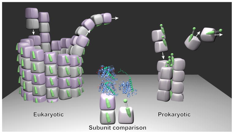
Figure 1.
Comparison of architecture for the three-stranded PhuZ cytomotive filament and the microtubule. (The figure is not intended to show a typical assembly/disassembly stage but a mixture, in order to display how the building blocks are added to or removed from the ends). The middle of the figure displays the eukaryotic tubulin dimer (on the left, PDB ID: 1JFF) and the PhuZ tubulin monomer (on the right, PDB ID: 3R4V). The C-terminal regions are marked in green. For eukaryotic tubulins polymerized in the microtubule, the C-terminal regions face outwards and are exposed to the environment. For PhuZ subunits polymerized in the three-stranded filaments, the C-terminal regions face inside and are responsible for both the intra-protofilament and inter-protofilament interactions. The tight or loose springs of the PhuZ tubulin subunits represent the compacted or extended forms, respectively.

