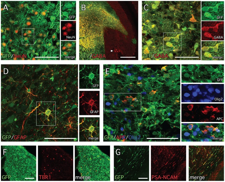
Figure 6. Neuronal phenotypes of grafted FA-iPS neural progenitors.
(A) The grafts were rich in differentiated neurons based on immunohistochemistry for GFP and NeuN. (B, C) Many of the neurons also expressed the neurotransmitter GABA (arrowhead in B shows host GABA+ Purkinje neurons). GFP+ cells with glial phenotypes were also present based on immunohistochemistry for (D) the astrocyte marker GFAP and (E) the oligodendrocyte markers Olig2 and APC. (F) A small population of GFP+/TBR1+ cells were scattered throughout the graft core. (G) The majority of the GFP+ cells with the morphology of migrating neuroblasts also expressed the cell adhesion molecule PSA-NCAM. Scale bars: A, C, D, E – 50 µm; B – 200 µm; F, G – 100 µm.

