
Figure 4. Conceptual application of multi-color STORM imaging using the same antibody species and same fluorophore.
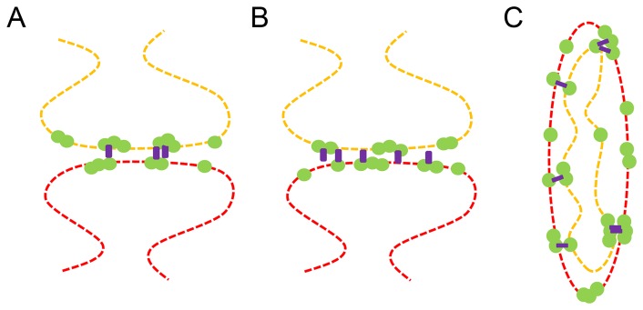
(A) Protein A (green) on the pre-synaptic structure (orange) and Protein B (green) on the post-synaptic structure (red) simultaneously colocalize with a third protein, Protein C (purple). (B) Protein C (purple) separately colocalizes with both Protein A (green) on the pre-synaptic structure (orange) and Protein B (green) on the post-synaptic structure (red) but rarely colocalizes with both proteins simultaneously. Sequential imaging using the same antibody species to label Protein A and B can be used to distinguish between these two scenarios (C) A second example following the same scenario as (A) and (B) but with mitochondrial inner and outer membrane proteins.