
Figure 7. Immunogold and osmium developed TEM reveals α-synuclein associated, intracellular fibrillar structures and increase of autophagosomes.
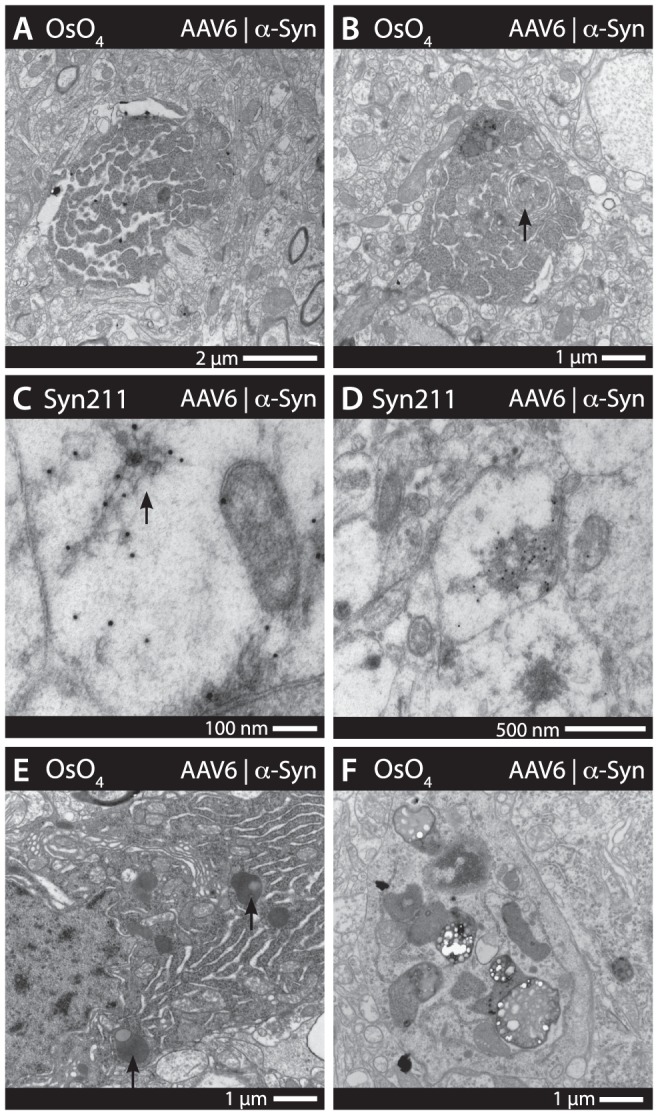
Additional electron microscopy revealed a number of pathological features, including apoptotic body cell remnants, present throughout the cerebral cortices (A). Autophagosomes forming from the Golgi (arrow) was a feature also present in cells with a high degree of apoptotic morphology (B). Human α-synuclein immunogold (syn211) staining revealed fibrillar-like structures (arrow in C) as well as larger inclusion bodies (D) present within the soma and axons (C–D). Subjectively, granular aggregates of lipofucins appeared to be a more common feature in α-synuclein expressing animals, suggesting lysosomal/proteosomal pathology. However, no absolute quantification was possible. (E–F).