
Figure 10. Morphological changes and evidence of α-synuclein uptake in IBA-1 positive microglia.
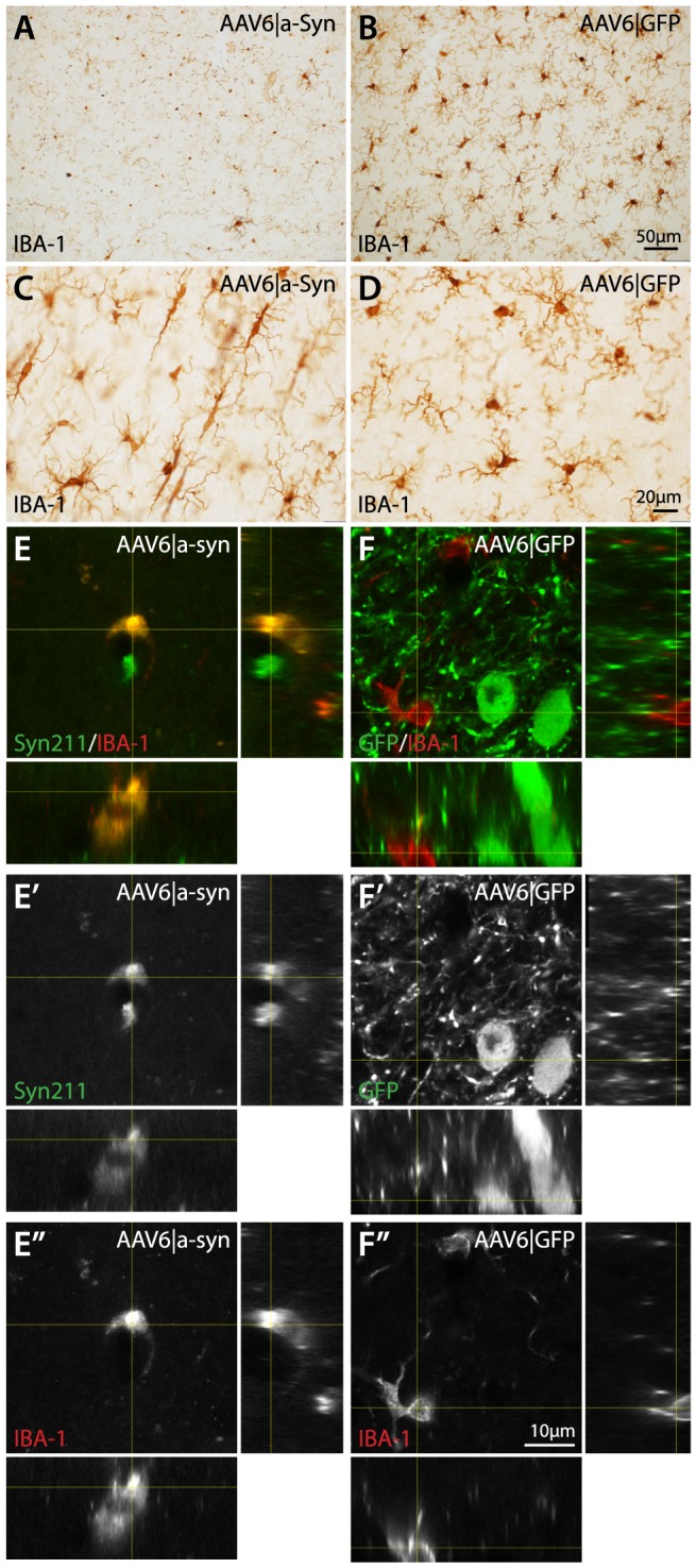
Immunohistochemical staining of IBA-1 using DAB precipitation coloring in the cerebral cortex revealed a substantial change in the microglial morphology in the cortex of AAV6|a-Syn animals compared to AAV6|GFP and Intact ctrl animals at 10 months post AAV injection (A–D). In the regions overlapping with the most intense human α-synuclein over-expression, most microglial cells appeared with very few visible processes and majority of the IBA-1 positive staining localized to the nucleus (A), this stood in stark contrast to the normal IBA-1 staining of microglia in the corresponding cortical regions of AAV6|GFP animals (B). In surrounding cortical regions, the microglial morphology in AAV6|a-Syn animals showed a second distinct morphology, with elongated cell body and fewer processer, suggesting a migratory profile (C). Using double-labeled, fluorescence with IBA-1 and human α-synuclein/GFP antibodies in a scanning confocal microscope, the presence of α-synuclein in microglia was investigated (E–F″). At the core of the human α-synuclein over-expression in the AAV6|a-Syn animals, microglia were found with significantly changes morphology and intra-cellular α-synuclein (E–E″). Interestingly, two localization patterns were observed, both in the cell soma in circular, vesicular structures and as small accumulations in distal processes of the microglia. No such patterns were observed in AAV6|GFP (F–F″) or intact controls.