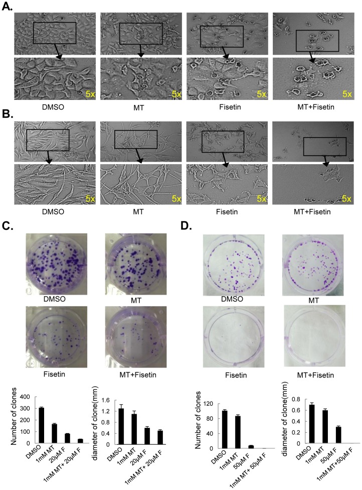
Figure 2. Melatonin enhanced fisetin-mediated cell morphology changes and clonal formation inhibitions.
(A–B), The cell morphology changes of MeWo (A) and Sk-mel-28 (B) cells treated with fisetin (F) (20 µM) and melatonin (MT) (1.0 mM) for 48 h were observed. The cells were photographed using a microscope with a magnification of 40×10 fold. (C–D), Clone formation in MeWo (C) and Sk-mel-28 cells(D) treated with fisetin (F) (20 µM) and melatonin (MT) (1.0 mM) for 48 h were observed.

