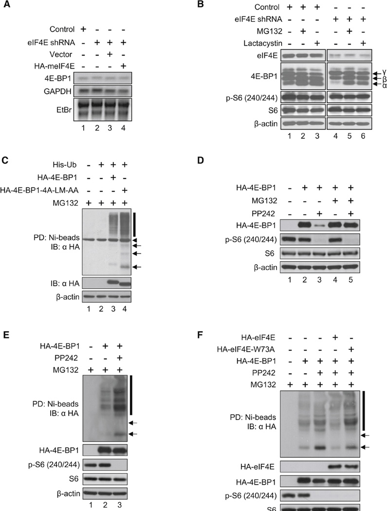
Figure 2.

Hypophosphorylated 4E-BP1 Is Degraded by the Ubiquitin-Proteasome Pathway. (A) 4E-BP1 mRNA levels in control and eIF4E-KD cells are shown. Total RNA was extracted from control, eIF4E-KD, and eIF4E-KD expressing the empty vector or HA-tagged meIF4E. 4E-BP1 mRNA levels were analyzed by northern blotting. GAPDH mRNA, 18S and 28S ribosomal RNAs detected by ethidium bromide (EtBr) staining served as loading controls. (B) Degradation of the hypophosphorylated 4E-BP1 is blocked by MG132 and Lactacystin. Control and eIF4E-KD cells were treated with DMSO, MG132 (20 µM), or Lactacystin (20 µM) for 3 hr. eIF4E, 4E-BP1 levels, and phosphorylated rpS6 were determined by western blotting. (C) eIF4E-unbound hypophosphorylated 4E-BP1 is highly ubiquitinated. 4E-BP1 and the eIF4E-unbound hypophosphorylated 4E-BP1 mimic mutant (4E-BP1-4A-LM-AA) were expressed in HEK293 cells together with His-Ub, and cells were treated with 20 µM MG132 for 6 hr. His-Ub proteins were pulled down with Ni-NTA agarose and analyzed by western blotting. Expression of exogenous proteins and β-actin in the lysates is shown in the panels below. Arrows and a bar indicate mono-ubiquitinated and polyubiquitinated 4E-BP1. An arrowhead indicates a nonspecific band. PD, pull-down; IB, immunoblot. (D) Hypophosphorylated 4E-BP1 levels are restored by MG132 treatment in cells treated with PP242. HA-4E-BP1 was transiently expressed in eIF4E-KD HEK293 cells, which were treated with 2.5 µM PP242 for 24 hr. Cells were cultured for the last 3 hr in the absence or presence of 20 µM MG132. 4E-BP1, rpS6, and β-actin levels were analyzed by western blotting. (E) Hypophosphorylated 4E-BP1 in PP242-treated cells is more ubiquitinated than in PP242-untreated cells. HA-4E-BP1 was expressed in 4E-BP DKO MEFs along with His-Ub, and cells were cultured in the absence or presence of PP242 for 24 hr, followed by in vitro ubiquitination assay. Pull-downed proteins were analyzed by western blotting. HA-4E-BP1, phosphorylated rpS6, and β-actin levels in cell lysates are shown below. Arrows and a bar indicate mono-ubiquitinated and polyubiquitinated 4E-BP1. (F) The interaction between 4E-BP1 and eIF4E prevents the hypophosphorylated 4E-BP1 from becoming ubiquitinated. HA-4E-BP1, HA-eIF4E, and HA-eIF4EW73A were expressed in 4E-BP DKO MEFs along with His-Ub, and an in vitro ubiquitination assay was performed as described in (C). See also Figure S1 and Table S2.

