
Figure 3.
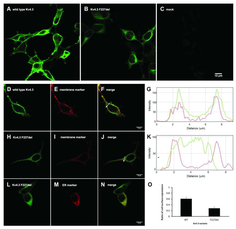
Confocal images demonstrating impaired cell surface expression and cytoplasmic retention of the mutant F227del Kv4.3. HEK-293T cells were transiently transfected with wild-type (A), p.F227del mutant (B) human Kv4.3 or empty vector (C) and immunostained with Kv4.3-specific antibody and green fluorophore-labeled secondary antibodies. Green fluorophore-labeled wild-type Kv4.3 (D) was expressed in the plasma membrane (E), colocalizing with DsRed-labeled membrane protein P0 (F). The spatial profile of fluorescent intensity of wild-type (wt) Kv4.3 (green) and P0 (red) along the arrow imposed on the images is shown in (G). The x axis displays the distance relative to the start point of the arrow and the y axis displays the fluorescence intensity. F227del Kv4.3 (H) was deficient in targeting to the plasma membrane (I, J). The spatial profile of fluorescent intensity of F227del Kv4.3 (green) and P0 (red) along the arrow imposed on the images is shown in (K). Instead, F227del Kv4.3 was retained in the cytoplasm (L) and colocalized with an ER-specific marker (M, N). The ratio of cell surface expression for the p.F227del Kv4.3 was significantly lower (O) than that observed in the wild type Kv4.3 (mean ± s.e.m.; p.F227del: 0.28 ± 0.04, n = 10; wild type Kv4.3: 0.61 ± 0.06, n = 10; p = 0.0014, Paired t test). Scale bar: 10 μm.