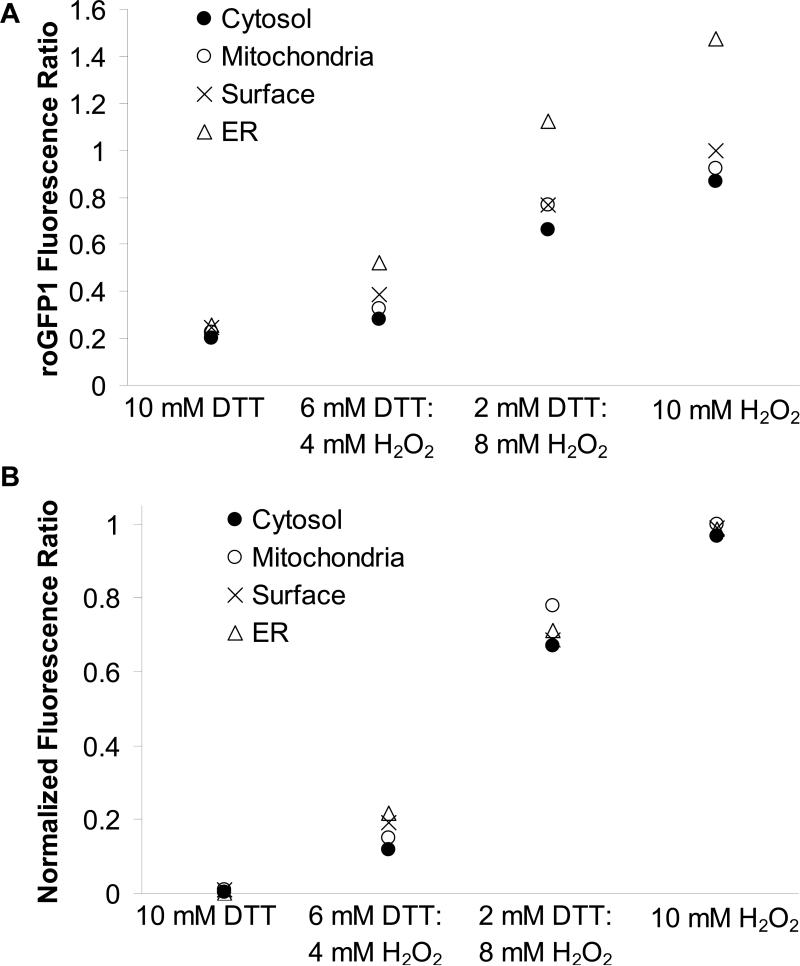
Fig. 3. Fluorescence properties of organelle-roGFP1.
A. CF15 cells expressing cytosolic, mitochondrial, ER and cell surface roGFP1 were treated with solutions containing DTTred and H2O2 at varying ratios (final concentration 10 mM in Ringer's). 385/474 nm fluorescence ratios for each compartment were recorded and background-subtracted (● cytosol, ○ mitochondria, × surface and ▲ ER; mean of n = 6 experiments shown, respectively), B. Normalization of data in Fig. 3A to maximal reduction and oxidation of roGFP1 fluorescence ratios show redox-sensing properties of roGFP1 were very similar for each organelle roGFP1.

