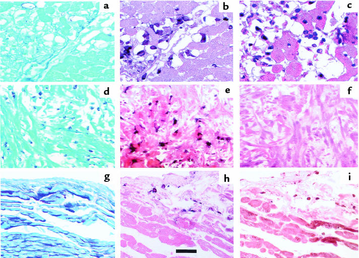
Figure 2.
Serial sections of ischemic myocardium collected 3 hours after reperfusion (a–c) and 5 hours after reperfusion (d–f). Control, nonischemic myocardium, documented by regional myocardial blood flows (g–i). Sections a, d, and g are stained with periodic acid-Schiff to demonstrate the presence of glycogen in normally perfused myocardium (g) and the absence of this substance in infarcted myocardium (a and d). Sections b, e, and h were stained with the AM-3K macrophage-specific antibody, and sections c, f, and i were stained for VLA-5. Note that 3 hours after reperfusion, monocytoid cells remain VLA-5+ for the most part, but after 5 hours, no VLA-5+ cells can be found in ischemic myocardium rich in AM-3K+ macrophages. In the control tissue, the few monocytes/macrophages present stain for VLA-5. Scale bar: 100 μm.

