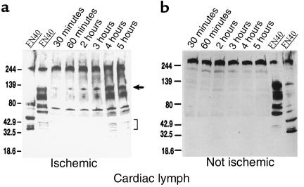
Figure 4.
FN and FN fragments in cardiac lymph. (a) Cardiac lymph from a dog with ischemia-reperfusion injury. Lanes 1 and 2 (positive controls) show immunoblots of human plasma FN enriched for 40- and 120-kDa FN fragments, respectively. Lanes 3–8 show lymph collected 30–300 minutes after reperfusion. An arrow indicates 120-kDa FN fragments. Brackets indicate smaller FN fragments found in lymph 4–5 hours after reperfusion. (b) Cardiac lymph from an operated dog that, because of good collateral circulation, did not experience cardiac ischemia (lanes 1–6). Almost all FN remains in the native 220-kDa form. Lanes 7 and 8 show human FN enriched for 120- and 40-kDa FN fragments, respectively, to show that FN fragments could have been detected under the conditions of this experiment.

